What is a Vascular Ultrasound scan?
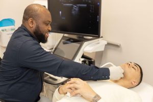
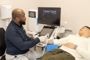
Using sound waves, a vascular ultrasound exam looks at the blood flow in your veins and arteries. High-frequency sound waves are transmitted from a handheld device, called a probe or transducer, that is placed on the body and moved around the area of interest. A warm water-based gel is applied to the skin to aid in the movement of the transducer. The transducer collects the sounds that bounce back and our Ultrasound machine then uses those sound waves to create an image.
Ultrasound exams are noninvasive, don’t use radiation, and images are captured in real-time, showing the movement of your blood flowing through your blood vessels.
Why I might need
this scan?
Detect Blood clots
Enlarged artery
Angioplasty surgery preparation
Varicose veins
Identify blockages and abnormalities
This scan can demonstrate
the presence of:
Deep venous thrombosis
Varicose veins
Venous reflux disease
Abnormal Aortic aneurysm
How to
prepare?
Depending on the location of the region, you may need to remove some clothing and wear a hospital gown.
A warm lubricating gel is applied to your skin to allow the probe to move smoothly.
No further preparation is required for this scan
What do I get with
this scan?
A radiologist’s written report of the findings will be provided to your referring practitioner, with images where necessary, to demonstrate abnormal findings.


